




This content was first published on IGR – Italian Gemological Review no. 22 in 2026. The information provided here is therefore current as of the original publication date.

Photomicrography has always been one of the most important aspects of gemology. Through the microscope it is possible to observe and document the internal world of gemstones, making visible details that are fundamental for study, identification and communication.
How has gemological photomicrography evolved from the analog era to the digital age? What is its role in education? As imaging techniques improve, the educational role of the image becomes stronger, photographic databases are created, and new forms of communication emerge on social media alongside exhibitions and dedicated competitions. One issue remains central: finding the right balance between scientific documentation and aesthetic expression.
Evolution of Photomicrography: From Film to Digital
Since the early twentieth century, photomicrography has played a central role in the development of modern gemology. The first images of inclusions, recorded on early photographic plates in the nineteenth century, date back to 1834 and were produced by Sir David Brewster. This marked the beginning of the systematic use of the microscope in gemology for the study of inclusions and it became an indispensable means of documenting and sharing observations that could not be described by words alone.
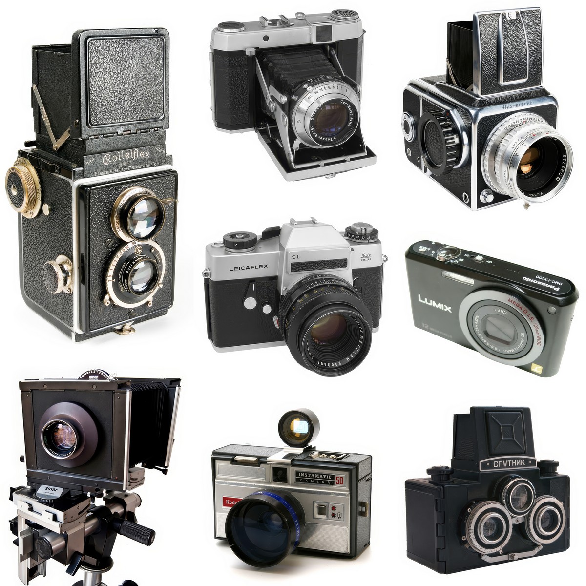
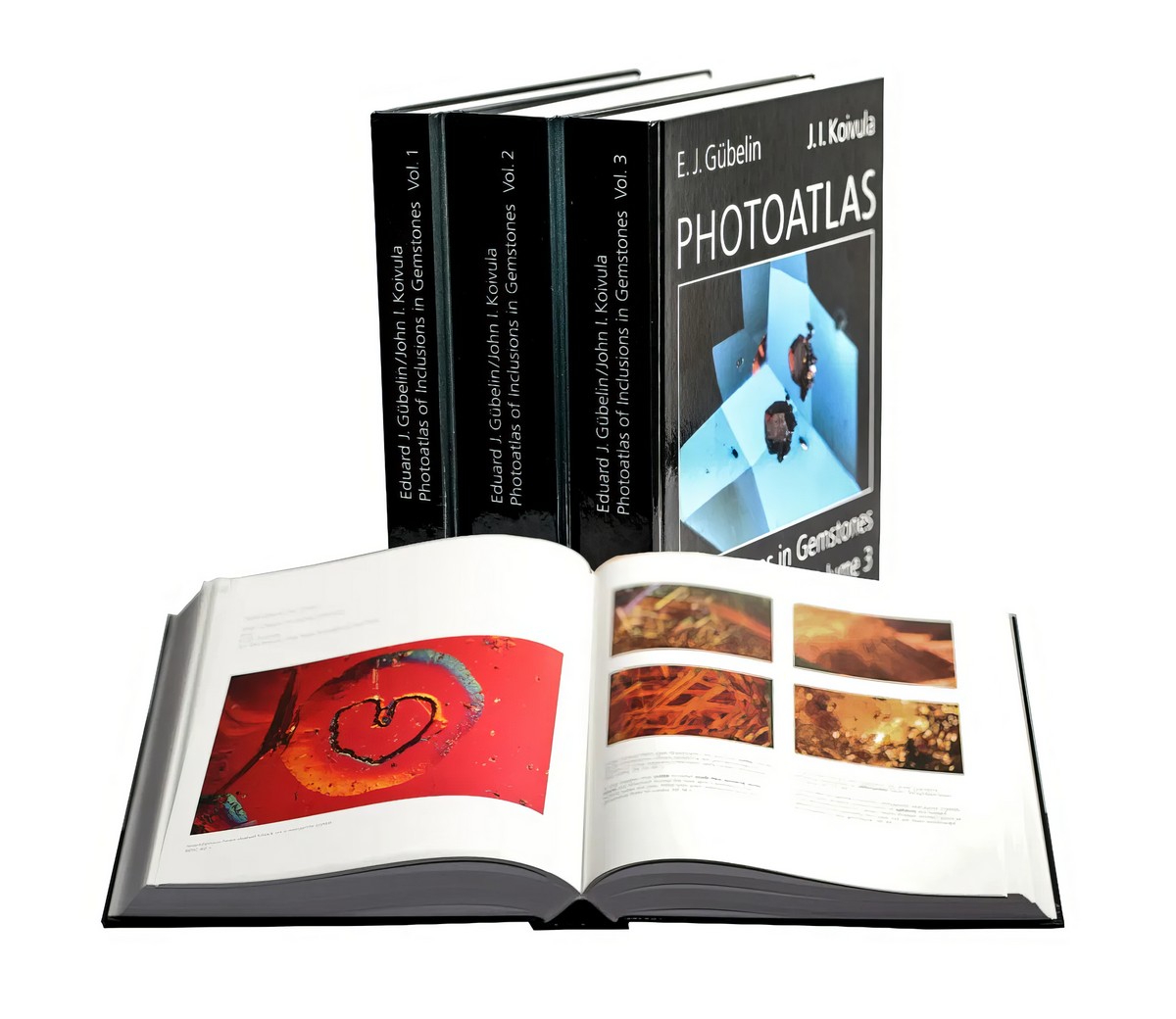
From around 1890 film cameras using gelatin silver bromide emulsions became common and improvements in sensitivity and portability represented a real turning point. Until the 1940s bellows cameras were used and these were not very versatile and difficult to connect to microscopes. In the 1970s more practical models appeared and during that period the idea of the PhotoAtlas volumes was born, destined to become a fundamental reference for generations of gemologists (Figure 1).
Working in the analog era meant having very limited control over exposure parameters. Microscope adapters did not allow precise focus control and focusing took place only through the viewfinder without any electronic support. John Koivula and Eduard Gübelin, absolute pioneers in the field, created extraordinary images for their PhotoAtlas of Inclusions in Gemstones volumes 1, 2 and 3 despite these limitations. Entire rolls of film were used in the hope of obtaining a single valid image and long waiting times for development often ended in disappointment when the results were unusable (Figure 2).
Analog practice therefore presented clear limitations such as long processing times, high costs, difficulties with framing and exposure and problems with color rendering. Yet the images of the PhotoAtlas remain “heroic” even today. They were obtained with enormous effort and they remain scientifically sound and surprisingly effective. Much like the first Disney animated films, produced with artisanal methods yet still capable of inspiring admiration.
Beyond the images themselves, the lasting value of the PhotoAtlas lies in the mineralogical notes that accompany them, written on the basis of deep geological and crystallographic knowledge. At that time diagnoses relied exclusively on these parameters. Today confocal micro Raman spectroscopy allows spectral analysis of individual inclusions with high precision. The technical quality of photography has improved over time, but the fundamental gemological principles remain unchanged.
The Educational Role of Photomicrography
The lesson left by Koivula and Gübelin is clear. Photomicrography was born with a primarily educational objective. Even today the microscopic image is an irreplaceable tool in gemological training because it allows the documentation of phenomena that are invisible to the naked eye and supports understanding of their complexity.
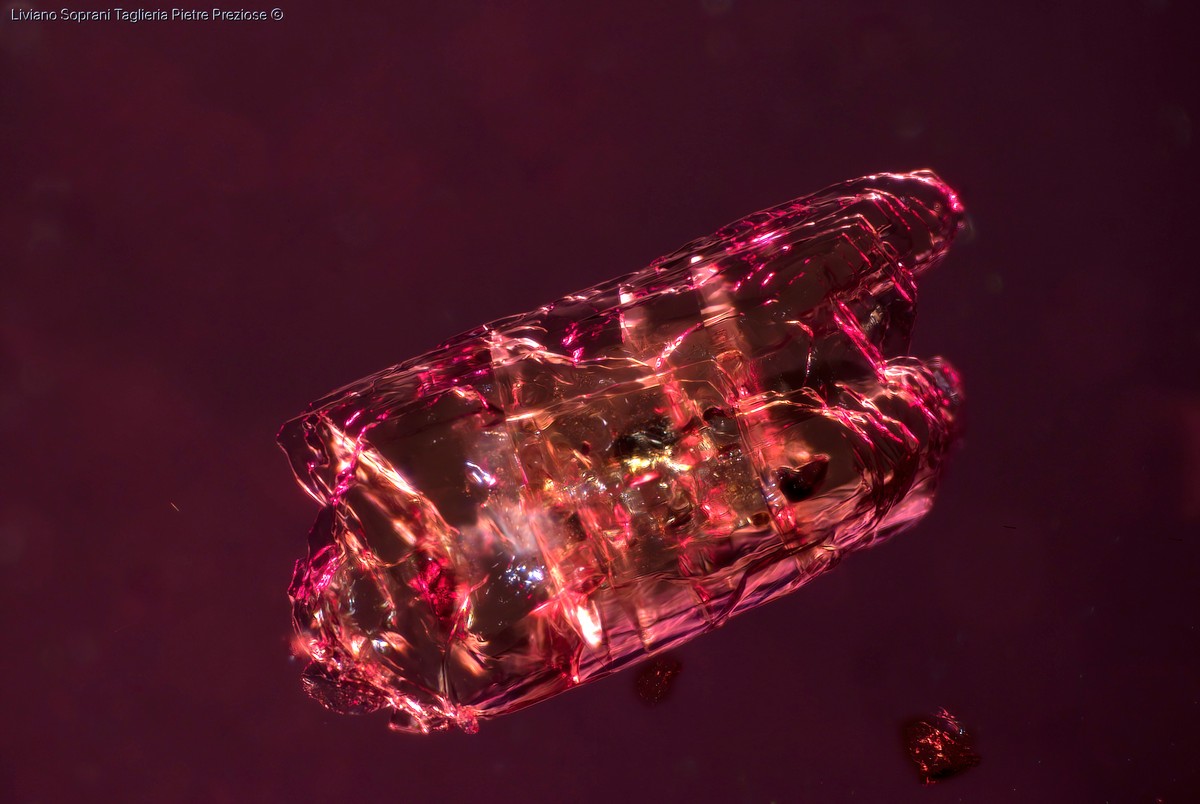
The study of inclusions, whether solid, liquid or gaseous, helps reconstruct the genesis and origin of a gemstone and helps identify possible treatments and the extent to which they have modified and improved its appearance.
Natural stones show irregular inclusions, fractures, strain halos, color zoning, twinning or growth structures and minute crystals linked to geological processes (Figure 3).
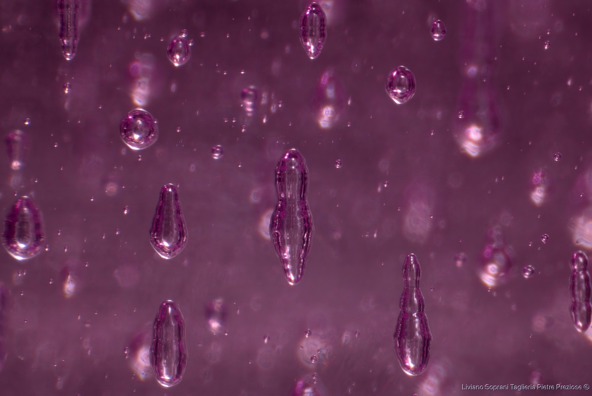
Synthetic stones instead display features typical of artificial production such as curved color or growth zoning and perfectly spherical or elongated gas bubbles and they may also contain flux residues or metallic platelets from contact with the crucible (Figure 4).
Treated stones may reveal colorless or colored filling substances, chromatic alterations or signs of diffusion, partially healed liquid feathers, dissolved crystals and strain halos caused by heating as well as surface coatings or laser drill holes (Figure 5).
Photomicrography allows these differences to be documented in a permanent and objective way and becomes a fundamental resource for teaching and for the creation of reference archives.
In recent years extensive photographic databases have developed both within laboratories and online. Comparing images of inclusions from gemstones of different geographic origins allows the development of diagnostic sensitivity that no theoretical text can replace.
In an era increasingly dominated by instrumental and spectroscopic data, photomicrography continues to stimulate direct observation and deductive reasoning. Thanks to digital technology every student can build a personal visual library that is useful for study, comparison and future professional practice.
Digital Imaging, Stacking and Post-Production
The widespread diffusion of photomicrography is linked to the advent of digital photography which completely changed the previous paradigm. Modern cameras equipped with increasingly high performance sensors make it possible to obtain high quality images in real time.
The possibility of checking the result even before taking the photograph allows correction of framing, exposure time, ISO and focus. In addition, computers connected to cameras allow image processing and large scale archiving. All this has made photomicrography accessible to an ever wider audience.
A fundamental contribution has come from focus stacking techniques which overcome the limits of shallow depth of field. By capturing multiple images at different focal planes and merging them later with dedicated software, it is possible to obtain a photograph with extended depth of field that is sharp in every part.
In the analog era the gemologist had to choose which portion of the inclusion to focus on and inevitably gave up other details. Today stacking allows an inclusion to be documented in its entirety.
The real turning point of digital photography, however, lies in post-production editing. Through it one can optimize contrast, correct brightness, adjust color saturation and vibrance, increase sharpness and correct white balance in order to achieve a result that is as faithful as possible to what is observed under the microscope. The camera does not always reproduce exactly what the eye perceives in live observation and this depends on sensor quality and lighting conditions.
The possibilities offered by image processing software are enormous and they imply a precise ethical responsibility. The gemologist must decide whether to adhere strictly to scientific accuracy or to favor a more evocative and artistic result. Excessive retouching may produce visually spectacular images that are scientifically misleading.
Each photomicrographer has a personal style and philosophy. I personally try to maintain gemological authenticity and avoid extreme retouching, color alterations or effects that may remove or falsify information and distance the image from what I observe through the eyepieces. Photography should remain descriptive, educational and informative in line with the spirit of the PhotoAtlas, although everyone is free to choose whether to remain within this context or move toward a more artistic and emotional approach.
Between Documentation and Visual Impact
Today photomicrography spreads rapidly thanks to social media where students and professionals share images of reference inclusions, unusual features or particularly evocative subjects. For scientific documentation purposes image manipulation should be limited to what is strictly necessary to highlight real phenomena.
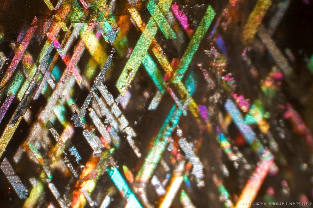
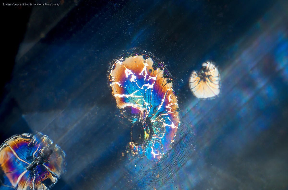
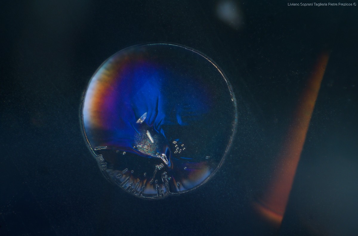
In competitive or outreach contexts the public tends to favor images with strong visual and emotional impact. Iridescence, for example, immediately captures attention even though it often offers limited diagnostic content. On social media the most spectacular images generally receive more attention than technically more complex ones (Figures 6a and 6b).
In laboratory routine one often encounters inclusions worthy of photographic documentation but time constraints do not always allow this. A high quality photomicrograph requires a long and complex process including identification of the optimal sample position for ideal framing, selection of the most suitable illumination to enhance the subject, numerous attempts with homemade diffusers or colored filters and careful management of incident light. Even in gemological competition juries it is not uncommon for emotional impact to prevail over technical and descriptive aspects.
In the future it would be appropriate to distinguish more clearly between scientific and artistic images and to attribute to each full legitimacy and specific purpose.
Instruments, Microscopes and Illumination
The quality of a photomicrograph largely depends on the instruments used. Today the gemologist can choose from a wide variety of cameras and microscopes and adapt equipment to the needs of analysis, research, documentation or communication.
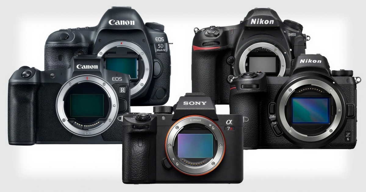
Digital single lens reflex cameras and more recent mirrorless models are the preferred choice for those seeking full control of shooting parameters and maximum image quality. They offer large sensors, the ability to work in RAW format and compatibility with a wide range of macro lenses and microscope adapters (Figure 7).
In recent years high end smartphones have also made enormous progress. When combined with digital or stereoscopic microscopes through appropriate supports they allow surprisingly good images in relation to cost and make photomicrography more accessible than ever.
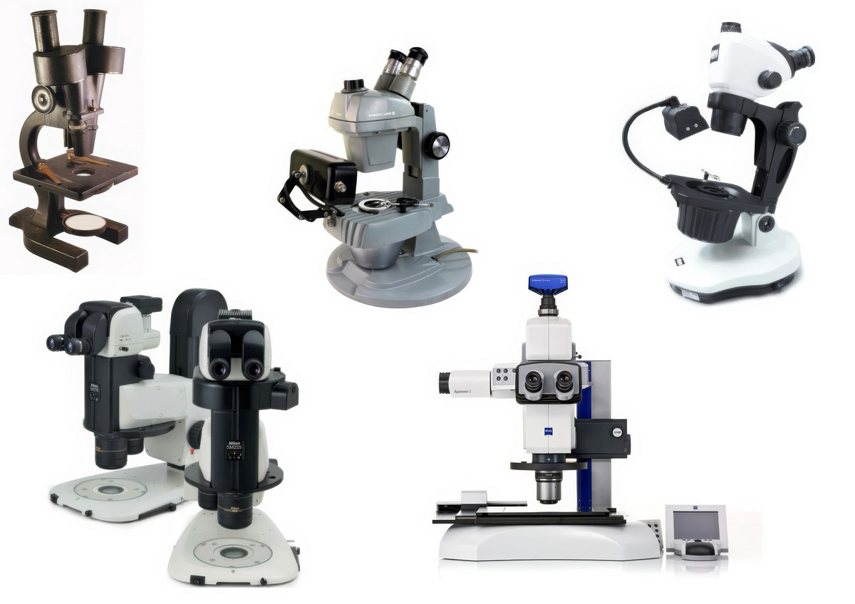
The gemological microscope, the heart of the entire system, may be optical or digital. Stereoscopic models with darkfield and transmitted light remain the most common in laboratories for three dimensional observation of inclusions (Figure 8).
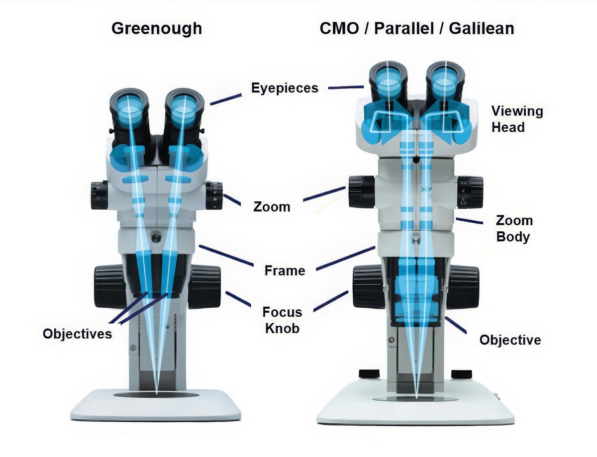
In optical stereo zoom microscopes it is necessary to distinguish between Greenough and CMO or Galilean architectures. The first consists of two independent converging optical systems that provide three dimensional vision similar to human binocular sight. Magnification rarely exceeds 80× and maximum resolution is below 450 line pairs per millimeter (Figure 9).
The second type, which is more expensive, uses a common large diameter interchangeable objective and two parallel optical paths that converge into the eyepieces. This allows magnifications up to 300× and resolution up to 1100 line pairs per millimeter although depth of field is drastically reduced compared to Greenough systems.
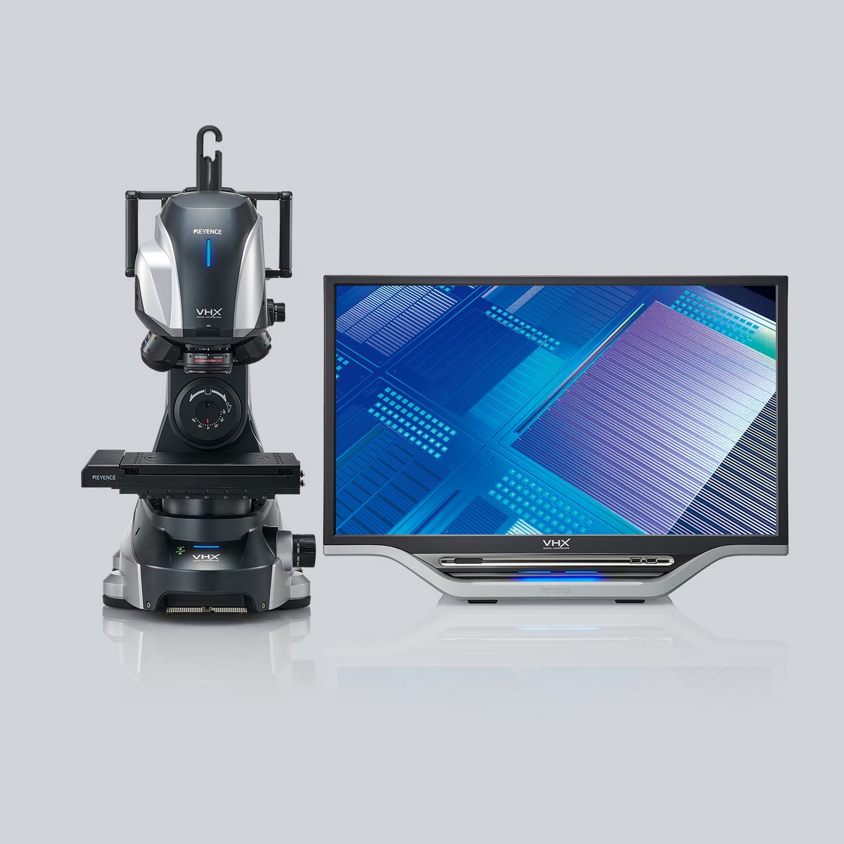
Alongside traditional optical instruments integrated digital microscopes are becoming increasingly common and in these systems the camera is part of the instrument body. These systems often include acquisition software and automatic stacking functions and they offer a simplified workflow and immediate results that are ideal for teaching laboratories and small research centers. Image quality, however, remains lower than that obtainable with optical microscopes and full frame cameras (Figure 10).
Whatever type of microscope is used control of illumination is fundamental. The type of light chosen, the direction of incident light, intensity and color temperature all strongly influence the final result. Halogen or LED light sources, auxiliary fiber optic illuminators, LED ring lights and diffused lighting systems allow selection of which details to highlight in inclusions and diffusers make it possible to reduce unwanted reflections by modulating light intensity.
Lighting techniques require experimentation and advanced knowledge. Fiber optic illumination, neon lighting and darkfield all introduce a certain degree of subjectivity similar in concept to that discussed for post-production.

In any case the organization of the photographic setup expresses the photographer’s point of view and each choice reflects intention. The photomicrographer may use diffusers when fiber optic light produces excessive illumination. Colored filters may be placed between the light source and the subject to create a more emotional atmosphere or to emphasize a specific color. A good example is my image that won the Gem-A Photographer of the Year 2024 competition. I used a red background placed between the darkfield base and the gemstone. Instead of relying on simple post-production retouching I chose a chromatic effect that was already present at the moment of capture in order to increase contrast and atmosphere. This made the image less realistic but at the same time it gave greater prominence without distortion to the hematite inclusion that I wanted to emphasize and it enhanced a color that was already present. In my view this was a balanced and appropriate choice because the theme of that competition was more artistic in nature (Figure 11).
The Artistic Dimension of Gemological Photomicrography
While maintaining solid scientific and educational value gemological photomicrography has now gone beyond simple documentation. The interior of gemstones reveals a microcosm of forms and colors that recall landscapes abstract structures and compositions of strong visual appeal.
Inclusions move from being purely diagnostic elements to becoming subjects that can enter into dialogue with painting and art history. In this direction stands the exhibition “Frammenti di meraviglia” (Fragments of Wonder), which I curated in Naples as an initial reflection on authenticity of representation and the emotional power of gemological images. Color zoning chromatic effects intersecting and oriented lines act as a mirror of certain paths of twentieth century abstract painting.
Within the detail of an inclusion the gemologist discovers not only technical information but also a form of natural beauty that speaks directly to the eye. At this meeting point between scientific rigor and aesthetic sensitivity gemological photomicrography continues to renew itself and to inspire.
Article by Liviano Soprani, published on IGR – Italian Gemological Review #22 – Spring 2026.


