This content was first published on IGR – Italian Gemological Review no. 6 in 2019. The information provided here is therefore current as of the original publication date.

Introduction
Generally, diamonds are known in gemmology for the features related to their commercial value. In the last few years, thanks to the studies of a scientific team, including an Italian scientist, the professor F. Nestola of Padua University, diamonds have assumed a role of fundamental importance for the mineralogical and petrographic characterization of earth deep areas (Pearson et al. 2014).
The team of scientists carrying out this research studies natural diamonds in its raw state, describing the morphological, optical and surface features and characterizing the fluid and solid inclusions.
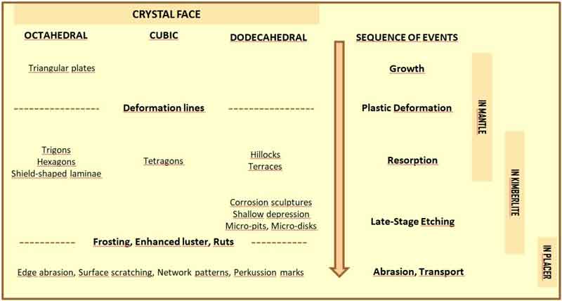
The study of the morphology of the diamonds provides important information on petrogenetic processes that have undergone these minerals, including their source from primary deposits or from secondary deposits (placers).
Natural diamonds occur as single crystals, as aggregates of a few crystals, or as cluster of countless small crystallites. The different morphologies (shape, size, texture surface, number of crystals, etc.) reflect the growth conditions and provide valuable information about the processes that led to the diamond formation. The classification and interpretation of the morphologies of diamonds has been a matter of debate for more than a century. Initial studies of diamonds were mostly descriptive, and they often focused on diamonds from a specific deposit or region. More comprehensive diamond classifications were subsequently developed. The classification scheme of Orlov (1977), for example, distinguishes eleven types of diamonds (varieties I-XI) based on differences in their morphology, internal structures, physical properties and impurities (Erlich et al. 2002). Other, more generalized diamond classification schemes emphasize the growth mechanism that produce the different morphological types of diamond. In Tappert & Tappert 2011, three principle types of diamond are distinguished: mono crystalline diamonds, fibrous diamonds and polycrystalline diamonds; this classification is based on differences in morphology and growth mechanism.
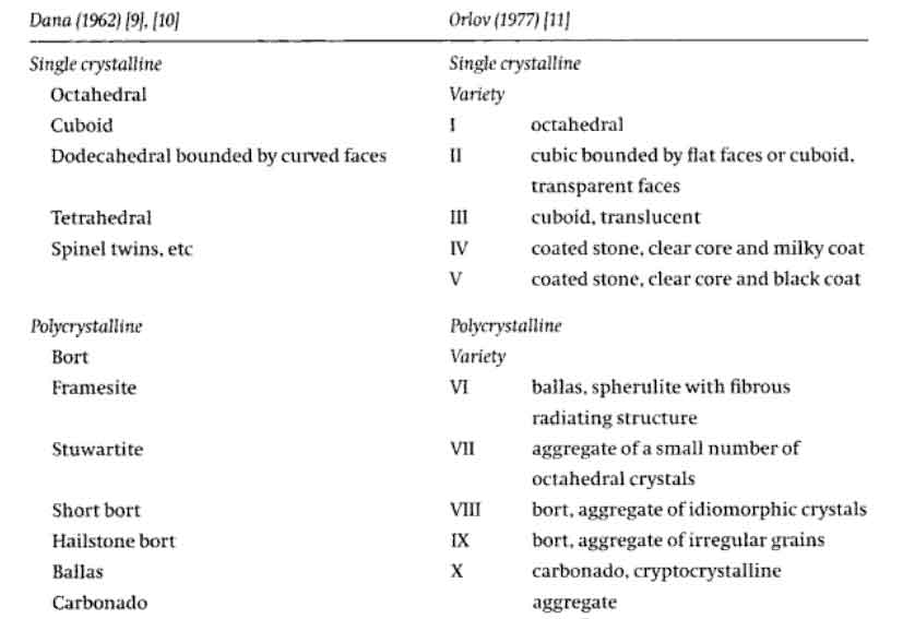
This work is focused exclusively on morphological description of natural, uncut diamonds (crystal shape, colors and surface texture) coming from Real Museo Mineralogico of Centro Musei delle Scienze Naturali of the Univeristy of Naples Federico II.
Materials and method
Morphological analyses were performed with Zeiss V20 stereomicroscope, with the Zeiss Axio Imager A1m petrographic microscope at reflected and transmitted light (DSC laboratory of Federico II University of Naples) and with Scanning Electron Microscope SEM-FEI Quanta 200 FEG (IPCB-CNR, Pozzuoli).
Results and Discussion
The Museum has 13 diamonds, 11 single crystals for a total 8.621 carats, and 2 crystals are on kimberlitic matrix. The diamonds were introduced in the Museum during the first half of 1800 with the exception of the sample with inventory number (N.I.) 2479, probably acquired by exchange in 1978. Six diamonds came from Brazil, six ones from Kimberly (South Africa) and one from Siberia, unfortunately we haven’t got more detailed information about their provenience. In particular, between the diamonds coming from Brazil, the samples A38, A39, A40, A41 and A42, there was part of the Collection Orittognostica of Matteo Tondi (1762-1835). Shown below the morphological description of some diamond collection of Royal Mineralogical Museum.
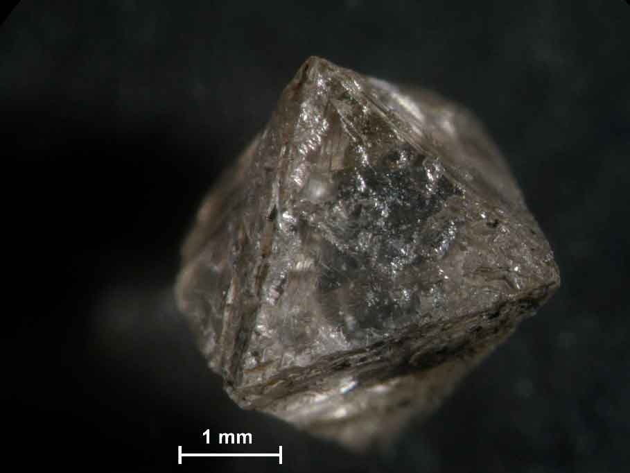
The F20 crystal coming from Brazil, is acquired by Dr. Adam August Krantz in 1851. The F20 grey diamond (0.12 ct) shows octahedral habit with deep etch-channels.
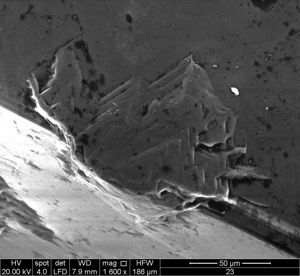
This diamond shows on its crystal faces some multiple imbricated triangular plates, with growth layer of variable thickness. Sometimes are observed multiple flat-bottomed trigons, oriented in the direction opposite to the underlying octahedral crystal face (negative trigons). The diamond also exhibits small elongated hillocks.
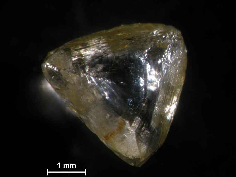
The N°23 crystal (0.04 ct), coming from Kimberley (South Africa) is a resorbed octahedral macle with pale yellow color. The diamond also exhibits brown irradiation spots. It is possible to observe the presence of a straight twinning line that marks the contact between the diamond crystals. A macle is an intergrowth of two flattened octahedral crystals that are connected along an octahedral plane with one of the crystals being oriented in the opposite direction.
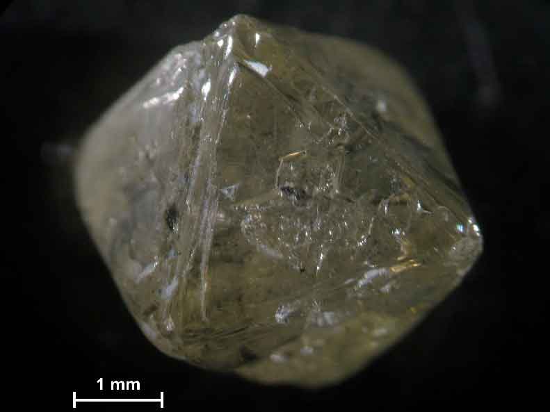
The N°62 diamond coming from Kimberley (South Africa) and is inserted in the collection of Museum by Prof A. Scherillo (director of Museum from 1939 to 1977) in 1967. The sample shows a partially resorbed octahedral habit, resorbed crystal edges and corners (5.99 ct). This diamond exhibits single and multiple triangular plates, shield-shaped laminae and triangular pits. On the surface of triangular faces it is visible the pyramidal, terraced and flat bottomed negative trigons (are oriented in the direction opposite to the underlying crystal face).
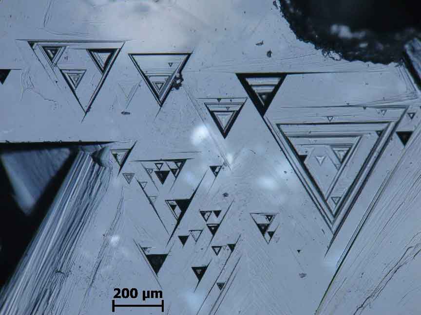
In particular negative trigons are considered to form when original growth surface is etched by fluid at high temperatures (~1000°C; Tappert et al. 2011).
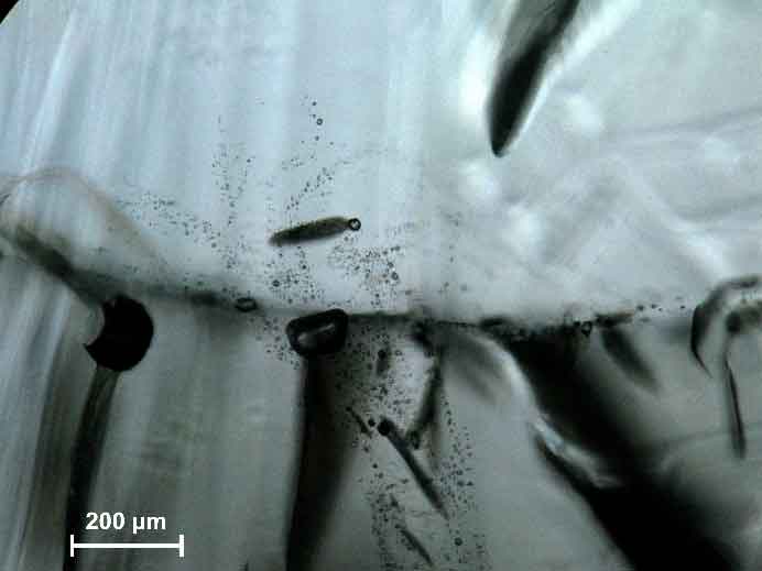
The N°62 diamond is characterized also by the presence of numerous inclusions sometimes associated with disc-shaped fracture system (Figure 8). The presence of inclusions conditions the color and clarity of a diamond, small particles of graphite or other minerals produce a grey appearance.
Diamond of Tondi Collection
Five diamonds (N.I A38, A39, A40, A41 and A42) was part of the Collection Orittognostica of Matteo Tondi (director of Museum from 1816 to 1835).
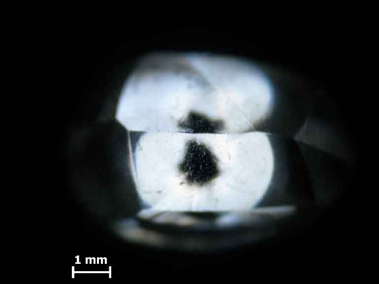
The A38 diamond (0.47 ct) is characterized by rounded dodecahedral habit, by presence of ancillar ridge and one black central inclusion.

The colorless and transparent diamond shows also the network patterns. On dodecahedral crystal faces, the intersection of two sets of grooves produces a distinctive rhombic pattern.
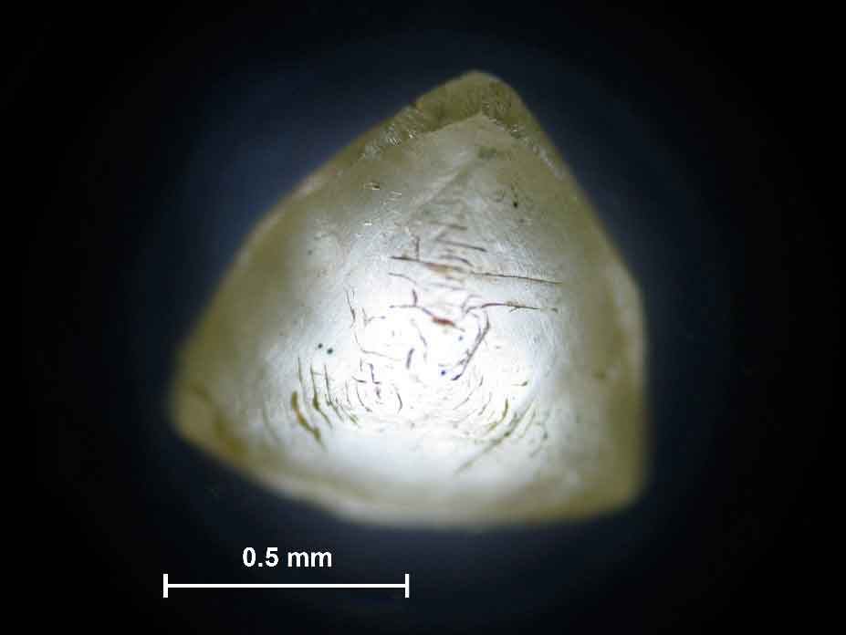
The A40 crystal (0.16 ct) is dodecahedral macle with yellow color with different intensities zones. It is possible to observe the presence of a straight twinning line that marks the contact between the diamond crystals. This diamond is characterized by the presences of green and brown single spots. The A40 diamond appears dull for the presences of hillocks groups. They are aligned concentrically around the three fold axes of dodecahedral crystal faces. The hillocks are drop-shaped and elongated and sometimes abraded. On the diamond surface are also visible the percussion marks. The fractures can extend deep into the interior of the diamond following the direction of crystal planes.
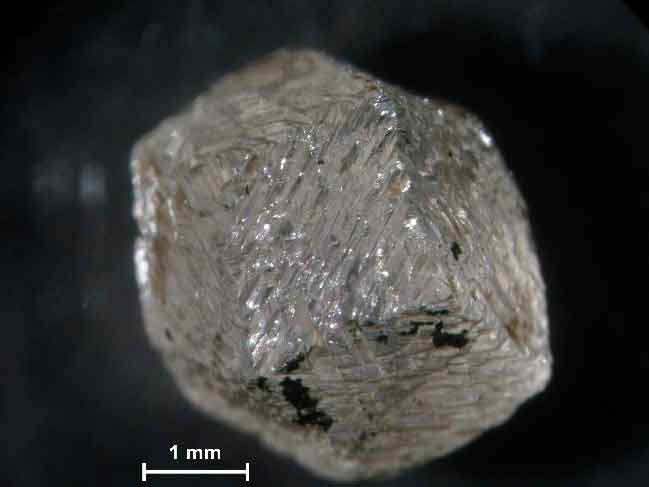
The A42 sample is a monocrystalline diamond (0.62 ct) with characteristic rugged crystal faces. This diamond shows octahedral habit in combination with cubic habit (cubo-octahedral habit; Tappert et al., 2011). This diamond is colorless, with yellow color zones. On the surface are visible impurities of black color.
Diamonds on matrix
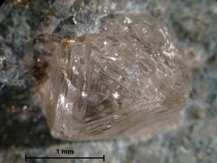
The N°2479 sample (3x2x2 cm) coming from North East of Siberia (Yakutia) and is introduced in the Museum in 1978. The N°2479 crystal is the colorless resorbed octahedral diamond on the kimberlitic matrix. The stepped octahedral diamond shows slightly resorbed rounded triangular growth plates and sometimes negative trigons, fracture and dark inclusions.
Conclusion
The diamonds studied are characterized by very different morphological features. They occur with monocrystalline (single crystals, twins) diamonds, with different shape (octahedral, dodecahedral, flattened and transitional), colors and a wide range of surface textures. The crystals are yellow, grey and colorless. The crystals exhibit triangular plates, trigons (terraced, flat-bottomed, and pyramidal), hillocks, terraces and etch-pits that usually causes the diamond to appear dull. Sometimes inclusion and extensive fracture system can be found in diamonds. This work has shown that, most probably, the acquisition of samples was aimed at deepening on studies of morphological crystallography. In effect the diamonds are characterized by different morphological and textural features, topic of primary interest in the field of mineralogy always.
Acknowledgments
Thanks to the Director of the Museum Centre of Natural and Physical Sciences of Federico II University, Prof. Cappelletti P. for granting the use of the images of the samples studied and thanks to Dr. Petti C. for the availability shown to consult the ancient catalogues and to the use of the samples.
References
Erlich, E.I., Hausel, W.D., 2002. Diamond deposits: origin, exploration, and history of discovery. Society for Mining, Metallurgy, and Exploration, Inc., Littleton, CO, USA, 374 pp
McCallum, M.E., Huntley, P.M., Falk, R.W., Otter, M.L., 1994. Morphological, resorption and etch feature trends of diamonds from kimberlite populations within Colorado–Wyoming State Line District, USA. In: Meyer, H.O.A., Leonardos, O.H. (Eds.), Diamonds: Characterisation, Genesis and Exploration. Special Publication, vol. 1/B. CPRM, pp. 32–50
Nestola F., Alvaro M., Pearson D.G., Shirey S.B. (2016). The Nature of Diamonds and Their Use in Earth’s Study, Lithos, 265, 1-350
Orlov Yu. L. 1977. The Mineralogy of the Diamond. Geological Magazine 115(06), 474
Pearson D.G., Brenker F.E., Nestola F., McNeill J., Nasdala L., Hutchison M.T., Matveev S., Mather K., Silversmit G., Schmitz S., Vekemans B. Vincze L. (2014). Hydrous mantle transition zone indicated by ringwoodite included within diamond. Nature, 507, 221–224
Sunagawa I. (2005). Crystal: Growth, Morphology and Perfection. Cambridge University Press, United Kingdom 1-299
Tappert, R. & Tappert, M.C. (2011): Diamonds in Nature: A Guide to Rough Diamonds. Spinger-Verlag Berlin Heidelberg 1-142.
Article by Manuela Rossi* and Maria Rosaria Ghiara**, published on IGR – Italian Gemological Review #6, Spring 2019
* Department of Earth, Environment and Resources Sciences, University of Naples Federico II, Via Cinthia, 80126 Naples, Italy;
** Royal Mineralogical Museum, University of Naples Federico II, Via Mezzocannone 8, 80134 Naples, Italy; Corresponding author’s email address: manuela.rossi@unina.it