This content was first published on IGR – Italian Gemological Review no. 2 in 2017. The information provided here is therefore current as of the original publication date.

One of the essential requirements of gemological analysis is that the gemstones should not be damaged or permanently altered. In other words the analysis must be non-destructive. This perfectly understandable limitation prevents gemologists and gemological laboratories to use many techniques widely available for analyzing other kind of samples by sacrificing small parts of them. Raman spectroscopy is considered to be the best-known identification technique in material analysis as it is extremely reliable and, at the same time, fast and absolutely non-invasive for gemstones.
The Raman effect
When a substance is irradiated with light, a small portion of this radiation is scattered. In scattering a photon, which can be considered as a single moving piece of light, randomly changes its direction of propagation. This effect is called as elastic scattering and is also known as Rayleigh scattering effect.
A very small fraction (approximately 1 in 10 million photons) of the light scatters in a different way. In this so called inelastic scattering or Raman scattering something else than just direction of propagation changes too. In this case, there is an exchange of energy between the photon of the incident radiation and the molecule, which will reach an excited state. Part of this energy is used by the matter to start a vibrational motion. Subsequently, the matter moves back to the ground state by releasing a photon whose energy is missing the quota needed to ignite the vibrational motion. The result is that the light has changed its color.
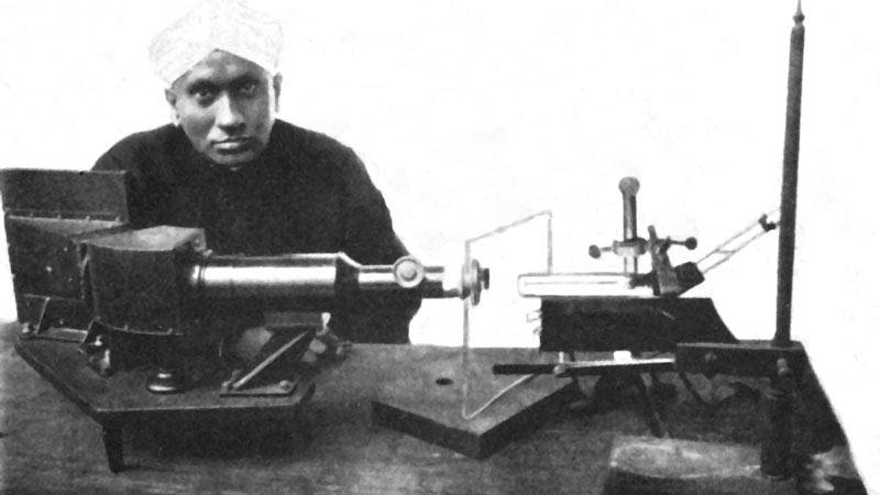
Raman scattering is closely related to the vibration modes of a substance; since these vibrations depend on both mass of the atoms, and the bonding forces & symmetries between them, it follows that the Raman spectrum of a substance represents a fingerprint which allows its unambiguous identification. For the discovery of this scattering, which took place in 1928, Sir C.V. Raman was awarded the Nobel Prize in Physics in 1930 (Figure 1).
An extraordinary aspect of this scattering, commonly referred to as Raman shift, is that it is not only unique and distinct for any given material but remains constant regardless the wavelength of used excitation source. This element is far from secondary if we think that in gemology, as in many other fields, we have to deal with a multitude of different materials which often react differently depending on the energy of the emission used to identify them. Some samples will in fact be easier to detect by using a source in the visible wavelength range, while for others that may exhibit luminescence reactions at these wavelengths, it may be necessary to use infrared units for excitation.
The Raman Spectrometer
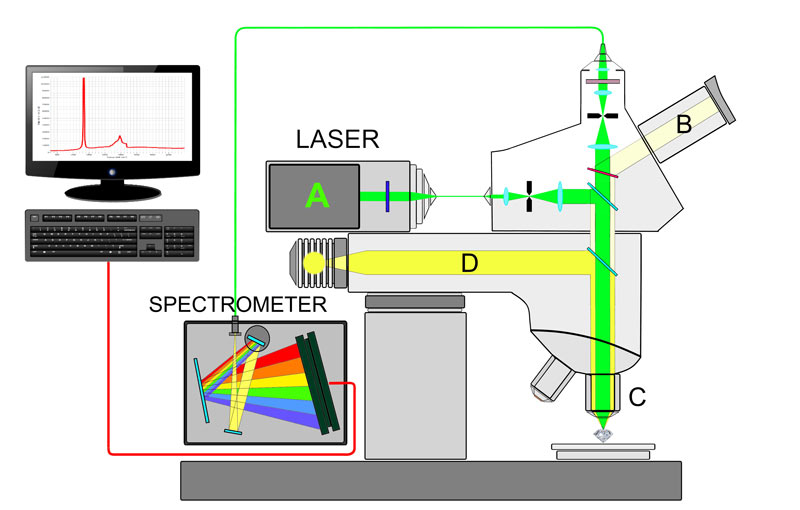
Conceptually the Raman unit it’s a quite elementary system: a strong monochromatic light source is focused on the sample. This emits a spectrum of radiation that is subsequently filtered so that only inelastic scattering reaches the detector. A computerized system will then transform the signal into a readable spectrum. If the schematic view of the system described is simple, in practice there are a number of problems that require highly advanced technical solutions. If we go back for a moment to the concept of inelastic scattering (caused by 1 photon out of 10 million) we can understand how small this is, to the limits of the detectable; It is therefore intuitive that, to increase its strength, it is necessary to have an extremely powerful monochromatic source. Until a few years ago the only system capable of meeting this requirement was the monochromator, an apparatus that, by means of diffraction gratings, disrupted the visible light in its monochrome components that could then be used to generate the Raman effect in the sample. It was a very expensive, cumbersome and barely efficient system. One of the two aspects that allowed the rapid spread of Raman spectroscopy in relatively recent times was the speed the LASERS have evolved. Thinking about it, given its characteristics, one might think that LASER was actually invented for Raman Spectroscopy: an extremely powerful monochromatic coherent beam available at a fairl affordable cost when compared to monochromator. The other factor that has greatly boosted the Raman technique is the adoption of Charge-Coupled Device (CCD) spectrometers, much more compact, cheap and in many cases even more efficient than the traditional Photomultipler Sensor Systems (PMT). The last critical aspect is the filters whose effectiveness, thanks to the progress in chemistry, has improved exponentially over the last decade.
Confocal MicroRaman
It is certainly worth briefly describing here a Raman apparatus which has led to a real turning point in gemology regarding the identification of inclusions in gemstones.
Until not many years ago, microscopic observation was the most commonly used method to identify the nature of inclusions. In combination with a microscopic and efficient lighting system, it was obviously necessary to have a deep understanding of the crystalline habits and appearance of the various elements and their possible associations with the gem analyzed. Due to the objective difficulties that this practice involved, however, the result remained, very often and with few exceptions, in the field of possibilities. The Confocal MicroRaman, although with few limitations, allows to identify inclusions within a gem with absolute certainty without damaging it. However, the process is not so simple as it seems and a powerful data processing system with specific software is required for the result to be appreciable. Inclusion then cannot be located too deep in the gem; In fact, the system records a spectrum that is the combination of the host material and the inclusion; if the first one is too overwhelming and/or superimposed, inclusion recognition can be extremely complicated if not impossible. In many cases, confocal MicroRamans have interchangeable Laser sources, usually visible and infrared, to broaden the range of available survey as much as possible.

Luminescence, annoying noise or precious resource?
It has been said that a wide range of wavelengths can be used to stimulate Raman diffusion, but what is the most effective for gemology? Raman systems can be grouped into two families, depending on whether the source is in the field of visible or infrared wavelength. Traditionally, the infrared emission (typically between 780 and 800 nm) has been the most preferred in the past: the reason behind this choice was to limit as much as possible an unwanted luminescence reaction in the samples. The most diverse chemical elements in the composition of the matter can react by generating an annoying emission, called as fluorescence, often in the visible range, which can completely or partially mask the very weak Raman diffusion making it impossible to be detected.
Emissions in the infrared region rarely cause this reaction and for this reason have long been preferred in the design of Raman systems. The negative side comes from the fact that very powerful lasers and extremely sensitive spectrometers are needed (the CCD sensors are barely efficient in the infrared) to obtain appreciable results. All of this leads to building costs that are normally three times higher than those of a Raman units that use visible light. The reason why, nowadays, the visible sources are gradually more preferred, is that any luminescence emissions, although in some cases can make it problematic or impossible to scan the Raman effect, provide us with additional and valuable information for Gemological analysis, not only for identifying materials but also to detect their possible treatments. Given the extraordinary importance of photoluminescence in the latest gemological studies, we will leave its discussion to a specific article that you can find in the next issue of the magazine.

Gemological applications
One of the conceptual pillars of gemology teaches us to collect as many data as possible – very often the detective’s image is used – before venturing a concrete hypothesis in identifying gems. Probably the most common and effective protocol envisages the gradual exclusion of candidates as diagnostic tests give their results, limiting the range to the lower number of possible options.
The higher the number of techniques used, the higher the ability to get the exact identification. Raman spectroscopy is probably the only system that, in the vast majority of cases, allows the fast and accurate identification of the gem – whether it is polished or rough – without the need for further confirmatory tests. This means, in practice, a drastic decrease in survey times to the benefit of the work of the gemologist. Some experts in the industry are releasing some criticisms based on the problematicness or even the inability to equip with Raman Spectroscopy, unless one has an academic degree in the subject. We apologize for not sharing this position. The operator does not necessarily need to be a scientist to achieve valid results. What you need is an understanding of the operating principle, the ability to compare results with a proven reliability database and, last but not least, the ability to see when the instrument is delivering incorrect or not completely reliable results, either for sample positioning problems, for its inadequacy, or for system malfunction. In practice, the operator is only required to correctly use the spectrometer and correctly compare the results with the database, no more no less what is done when the refractive index is taken: reading and comparing the values with R.I. tables. It is certainly useful to know the mathematical demonstration with which you come to the critical angle determination, but not for this reason the readings collected by an operator who is unaware of that will be less valid.
In gemology we are used to deal with a multitude of gems containing chromophore trace elements, the most famous being chromium. One of its peculiar properties is to emit a violent luminescence reaction if excited by certain high intensity visible radiations, just what happens when we want to collect a Raman spectrum. Rubies, emeralds, alexandrites, in many cases sapphires and other gems contain chromium in various percentages and thus detecting the Raman fingerprint is very often practically impossible. While this aspect might suggest a substantial uselessness of Raman spectroscopy for these materials, it is to be considered that photoluminescence reactions also characterize the various gems with extreme accuracy and can be recorded and appropriately compared by the same visible sources equipped Raman spectrometers if they have the appropriate spectral range. Typically, chromium emissions occur between 678 and 690 nm, sometimes just over 700 nm, and if our spectrometer has a range that includes these values, the analyzed gems may be identified by their photoluminescence spectra, even if the Raman spectrum is impossible to collect.
Synthetics, treatments and… contraindications
Very often, from the point of view of the results, the Raman spectrometer is conceptually and perhaps improperly compared to an “enhanced” refractometer. Indeed, as in the case of the latter, it provides no distinction between natural and synthetic gems. There are few but significant cases where the Raman fingerprint of natural and synthetic gems differs, one on all: the spinel. Other significant differences (using photoluminescence) can be found in emeralds, opals, impregnation treatments of jade and other gems. Metals do not produce any Raman signal or noise, and this facilitates the analysis of mounted gems.
Obviously as happens for all the instruments, Raman spectroscopy also has its Achilles heel: we know it gives its best for well-defined crystalline structures, whereas amorphous materials have some difficulty. Even black or very dark samples, by their own nature, tend to absorb their own Raman radiation, and that is why in many cases it is objectively complicated to obtain their readable spectra. On the other hand, the situation in which Raman spectroscopy demonstrates all its extraordinary gemological efficacy is related to non-”traditional” materials that are increasingly entering the market more and more frequently due to the chronic scarcity of the most traditionally used gems.
Whether identifying a necklace of phosphosiderite or magnesite can take relatively long time using traditional methodologies, for a Raman spectrometer it will be a matter of seconds. The possibility of analyzing any shape and size (less than 1 mm is enough), even rough, makes it the almost irreplaceable instrument in the modern gemological laboratory.
Conclusions
Thanks to latest progress in laser and miniaturization of CCD spectrometers, Raman spectroscopy is recently becoming a fast and effective single-gemologist analysis system, not being anymore an advanced tool available in few big gem laboratories. Its speed, safety and ease of use will contribute to its wider diffusion and we hope it will not take long before it will be included in the gemological courses as a basic tool for gem identification.
References
D. Bersani, P.P. Lottici, “Analytical and Bioanalytical Chemistry”, 2010 Aug. – 397 (7), 2631-2646.
R. Jasinevicius, “Characterization of vibrational and electronic features in the raman spectra of gem minerals”, MSc thesis, 2009, Department of Geosciences, University of Arizona, Tucson, 147p.
Article by Alberto Scarani and Mikko Åström, published on Rivista Italiana di Gemmologia #2, September 2017.